
副業バレないバイト短期日払いアルバイトのおすすめサイト
【裏ワザ】短期日払いOKな副業を即効で見つける方法とは!?

副業におすすめ!稼げる高収入バイト求人一覧
数ある副業アルバイトの中からおすすめの約50種類の求人をご紹介します!気になる業種を選んで自分にピッタリの高収入アルバイトを見つけてみてね!
●求人の給与、待遇、勤務時間、勤務地、募集要項などの詳細については『詳細を見る!』又は画像、お仕事のタイトルをクリックしてね。
●「今すぐ応募したい!」という方や「直接お仕事の説明や話を聞きたい!」という方は『今すぐ応募!』をクリックしてね。
分からないことがあったり、初めてで不安な方、どのお仕事にしようか迷っているは気軽にご相談ください♪女性スタッフがお話を聞いて、あなたに最適のお仕事をご案内します!
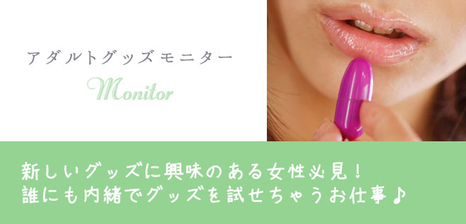
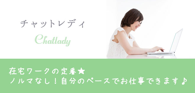
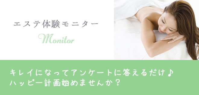
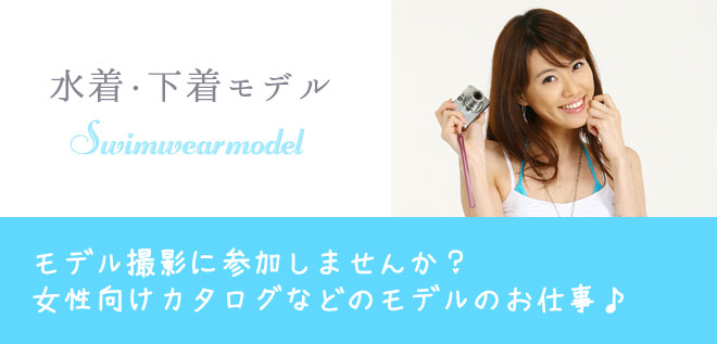
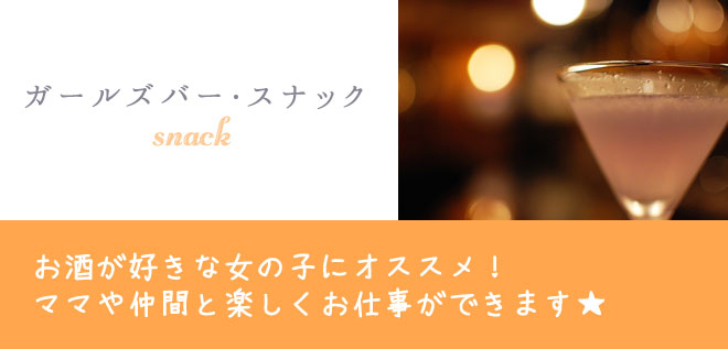
在宅ワーク系 ▼
平均日給 8000円~15000円以上
| オススメ度 | ★★★☆☆ |
|---|---|
| 応募資格 | 18歳以上(高校生不可) |
| 勤務エリア |
在宅勤務OK(スマートフォン又はパソコン必須) 【関西】 大阪・京都・兵庫・神戸・滋賀・奈良・和歌山 【関東】 東京・神奈川・横浜・千葉・埼玉・茨木・栃木・群馬 在宅勤務OK(スマートフォン又はパソコン必須) |
| 勤務時間 |
シフト 登録依頼制・週1日・月1日でもお仕事可能です! 勤務時間 24時間いつでもOK!(通勤の場合は要相談) |
| 待遇一覧 | 未経験・初心者OK!/高時給アルバイト/女性の高収入バイト/経験不問/資格不要/資格取得可能!/キャリアアップ/単発・短期・長期OK!/完全自由出勤制/新人保証あり/日給保証あり/時給保証あり/オープニングスタッフ募集/送迎あり/1日体験入店OK!/即日勤務可能/全額日払い/知人バレ・身バレ対策/給与交渉OK!/掛け持ちOK!/週1・月1~OK!/1時間から勤務可能/個人情報保護/専門学生・大学生歓迎!/派遣バイト/深夜/駅チカ/出稼ぎにもおすすめ!/その他 |
平均日給 4500円~25000円以上

| オススメ度 | ★★★★☆ |
|---|---|
| 応募資格 | 18歳以上(高校生不可) |
| 勤務エリア |
在宅勤務OK(スマートフォン又はパソコン必須) 【関西】 大阪・京都・兵庫・神戸・滋賀・奈良・和歌山 【関東】 東京・神奈川・横浜・千葉・埼玉・茨木・栃木・群馬 |
| 勤務時間 |
シフト 空いた時間にいつでもお仕事が可能です! 勤務時間 24時間好きな時間で販売可能!(店舗買取は要相談) |
| 待遇一覧 | 未経験・初心者OK!/高時給アルバイト/女性の高収入バイト/経験不問/資格不要/資格取得可能!/キャリアアップ/単発・短期・長期OK!/完全自由出勤制/新人保証あり/日給保証あり/時給保証あり/オープニングスタッフ募集/送迎あり/1日体験入店OK!/即日勤務可能/全額日払い/知人バレ・身バレ対策/給与交渉OK!/掛け持ちOK!/週1・月1~OK!/1時間から勤務可能/個人情報保護/専門学生・大学生歓迎!/派遣バイト/深夜/駅チカ/出稼ぎにもおすすめ!/その他 |
平均日給 18000円~46000円以上
| オススメ度 | ★★★★★ |
|---|---|
| 応募資格 | 18歳以上(高校生不可) |
| 勤務エリア |
在宅勤務OK(スマートフォン又はパソコン必須) 【関西】 大阪・京都・兵庫・神戸・滋賀・奈良・和歌山 【関東】 東京・神奈川・横浜・千葉・埼玉・茨木・栃木・群馬 在宅勤務OK(スマートフォン又はパソコン必須) |
| 勤務時間 |
シフト 空いた時間に1時間から、週1日・月1日でもお仕事可能! 勤務時間 通勤の場合は希望時間を選択可能・在宅の場合は24時間いつでもOK! |
| 待遇一覧 | 未経験・初心者OK!/高時給アルバイト/女性の高収入バイト/経験不問/資格不要/資格取得可能!/キャリアアップ/単発・短期・長期OK!/完全自由出勤制/新人保証あり/日給保証あり/時給保証あり/オープニングスタッフ募集/送迎あり/1日体験入店OK!/即日勤務可能/全額日払い/知人バレ・身バレ対策/給与交渉OK!/掛け持ちOK!/週1・月1~OK!/1時間から勤務可能/個人情報保護/専門学生・大学生歓迎!/派遣バイト/深夜/駅チカ/出稼ぎにもおすすめ!/その他 |
平均日給 3500円~12000円以上
| オススメ度 | ★★★★☆ |
|---|---|
| 応募資格 | 18歳以上(高校生不可) |
| 勤務エリア |
【関西】 大阪・京都・兵庫・神戸・滋賀・奈良・和歌山 【関東】 東京・神奈川・横浜・千葉・埼玉・茨木・栃木・群馬 |
| 勤務時間 |
シフト 登録依頼制・週1日・月1日でもお仕事可能です! 勤務時間 希望時間選択可・応相談 |
| 待遇一覧 | 未経験・初心者OK!/高時給アルバイト/女性の高収入バイト/経験不問/資格不要/資格取得可能!/キャリアアップ/単発・短期・長期OK!/完全自由出勤制/新人保証あり/日給保証あり/時給保証あり/オープニングスタッフ募集/送迎あり/1日体験入店OK!/即日勤務可能/全額日払い/知人バレ・身バレ対策/給与交渉OK!/掛け持ちOK!/週1・月1~OK!/1時間から勤務可能/個人情報保護/専門学生・大学生歓迎!/派遣バイト/深夜/駅チカ/出稼ぎにもおすすめ!/その他 |
モデルプロダクション系 ▼
平均日給 22000円~45000円以上

| オススメ度 | ★★★★★ |
|---|---|
| 応募資格 | 18歳以上(高校生不可) |
| 勤務エリア |
【関西】 大阪・京都・兵庫・神戸・滋賀・奈良・和歌山 【関東】 東京・神奈川・横浜・千葉・埼玉・茨木・栃木・群馬 |
| 勤務時間 |
シフト 登録依頼制・週1日・月1日でもお仕事可能! 勤務時間 希望時間選択可能・応相談 |
| 待遇一覧 | 未経験・初心者OK!/高時給アルバイト/女性の高収入バイト/経験不問/資格不要/資格取得可能!/キャリアアップ/単発・短期・長期OK!/完全自由出勤制/新人保証あり/日給保証あり/時給保証あり/オープニングスタッフ募集/送迎あり/1日体験入店OK!/即日勤務可能/全額日払い/知人バレ・身バレ対策/給与交渉OK!/掛け持ちOK!/週1・月1~OK!/1時間から勤務可能/個人情報保護/専門学生・大学生歓迎!/派遣バイト/深夜/駅チカ/出稼ぎにもおすすめ!/その他 |
平均日給 5000円~10000円以上

| オススメ度 | ★★★☆☆ |
|---|---|
| 応募資格 | 18歳以上(高校生不可) |
| 勤務エリア |
【関西】 大阪・京都・兵庫・神戸・滋賀・奈良・和歌山 【関東】 東京・神奈川・横浜・千葉・埼玉・茨木・栃木・群馬 |
| 勤務時間 |
シフト 登録依頼制・週1日・月1日でもお仕事可能! 勤務時間 希望時間選択可・応相談 |
| 待遇一覧 | 未経験・初心者OK!/高時給アルバイト/女性の高収入バイト/経験不問/資格不要/資格取得可能!/キャリアアップ/単発・短期・長期OK!/完全自由出勤制/新人保証あり/日給保証あり/時給保証あり/オープニングスタッフ募集/送迎あり/1日体験入店OK!/即日勤務可能/全額日払い/知人バレ・身バレ対策/給与交渉OK!/掛け持ちOK!/週1・月1~OK!/1時間から勤務可能/個人情報保護/専門学生・大学生歓迎!/派遣バイト/深夜/駅チカ/出稼ぎにもおすすめ!/その他 |
平均日給 28000円~50000円以上
| オススメ度 | ★★★☆☆ |
|---|---|
| 応募資格 | 18歳以上(高校生不可) |
| 勤務エリア |
【関西】 大阪・京都・兵庫・神戸・滋賀・奈良・和歌山 【関東】 東京・神奈川・横浜・千葉・埼玉・茨木・栃木・群馬 |
| 勤務時間 |
シフト 登録依頼制・週1日・月1日でもお仕事可能! 勤務時間 希望時間選択可・応相談 |
| 待遇一覧 | 未経験・初心者OK!/高時給アルバイト/女性の高収入バイト/経験不問/資格不要/資格取得可能!/キャリアアップ/単発・短期・長期OK!/完全自由出勤制/新人保証あり/日給保証あり/時給保証あり/オープニングスタッフ募集/送迎あり/1日体験入店OK!/即日勤務可能/全額日払い/知人バレ・身バレ対策/給与交渉OK!/掛け持ちOK!/週1・月1~OK!/1時間から勤務可能/個人情報保護/専門学生・大学生歓迎!/派遣バイト/深夜/駅チカ/出稼ぎにもおすすめ!/その他 |
平均日給 25000円~50000円以上

| オススメ度 | ★★★☆☆ |
|---|---|
| 応募資格 | 18歳以上(高校生不可) |
| 勤務エリア |
【関西】 大阪・京都・兵庫・神戸・滋賀・奈良・和歌山 【関東】 東京・神奈川・横浜・千葉・埼玉・茨木・栃木・群馬 |
| 勤務時間 |
シフト 登録依頼制・週1日・月1日でもお仕事可能! 勤務時間 希望時間選択可・応相談 |
| 待遇一覧 | 未経験・初心者OK!/高時給アルバイト/女性の高収入バイト/経験不問/資格不要/資格取得可能!/キャリアアップ/単発・短期・長期OK!/完全自由出勤制/新人保証あり/日給保証あり/時給保証あり/オープニングスタッフ募集/送迎あり/1日体験入店OK!/即日勤務可能/全額日払い/知人バレ・身バレ対策/給与交渉OK!/掛け持ちOK!/週1・月1~OK!/1時間から勤務可能/個人情報保護/専門学生・大学生歓迎!/派遣バイト/深夜/駅チカ/出稼ぎにもおすすめ!/その他 |
平均日給 32000円~60000円以上

| オススメ度 | ★★★☆☆ |
|---|---|
| 応募資格 | 18歳以上(高校生不可) |
| 勤務エリア |
【関西】 大阪・京都・兵庫・神戸・滋賀・奈良・和歌山 【関東】 東京・神奈川・横浜・千葉・埼玉・茨木・栃木・群馬 |
| 勤務時間 |
シフト 登録依頼制・週1日・月1日でもお仕事可能! 勤務時間 希望時間選択可能・応相談 |
| 待遇一覧 | 未経験・初心者OK!/高時給アルバイト/女性の高収入バイト/経験不問/資格不要/資格取得可能!/キャリアアップ/単発・短期・長期OK!/完全自由出勤制/新人保証あり/日給保証あり/時給保証あり/オープニングスタッフ募集/送迎あり/1日体験入店OK!/即日勤務可能/全額日払い/知人バレ・身バレ対策/給与交渉OK!/掛け持ちOK!/週1・月1~OK!/1時間から勤務可能/個人情報保護/専門学生・大学生歓迎!/派遣バイト/深夜/駅チカ/出稼ぎにもおすすめ!/その他 |
平均日給 100000円~100万円以上

| オススメ度 | ★★★★★ |
|---|---|
| 応募資格 | 18歳以上(高校生不可) |
| 勤務エリア |
【関西】 大阪・京都・兵庫・神戸・滋賀・奈良・和歌山 【関東】 東京・神奈川・横浜・千葉・埼玉・茨木・栃木・群馬 |
| 勤務時間 |
シフト 登録依頼制・週1日・月1日でもお仕事可能! 勤務時間 希望時間選択可能・応相談 |
| 待遇一覧 | 未経験・初心者OK!/高時給アルバイト/女性の高収入バイト/経験不問/資格不要/資格取得可能!/キャリアアップ/単発・短期・長期OK!/完全自由出勤制/新人保証あり/日給保証あり/時給保証あり/オープニングスタッフ募集/送迎あり/1日体験入店OK!/即日勤務可能/全額日払い/知人バレ・身バレ対策/給与交渉OK!/掛け持ちOK!/週1・月1~OK!/1時間から勤務可能/個人情報保護/専門学生・大学生歓迎!/派遣バイト/深夜/駅チカ/出稼ぎにもおすすめ!/その他 |
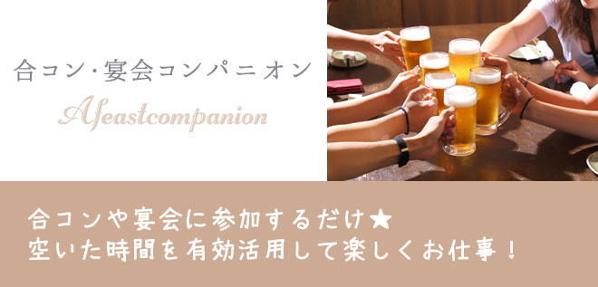
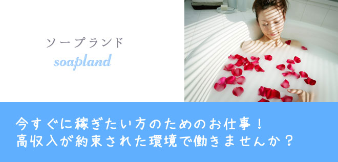
ナイトワーク系 ▼
平均日給 18000円~37500円以上
| オススメ度 | ★★★★★ |
|---|---|
| 応募資格 | 18歳以上(高校生不可) |
| 勤務エリア |
【関西】 大阪・京都・兵庫・神戸・滋賀・奈良・和歌山 【関東】 東京・神奈川・横浜・千葉・埼玉・茨木・栃木・群馬 |
| 勤務時間 |
シフト シフト自由で週1日からお仕事可能! 勤務時間 営業時間内から自由に選択可能・応相談 |
| 待遇一覧 | 未経験・初心者OK!/高時給アルバイト/女性の高収入バイト/経験不問/資格不要/資格取得可能!/キャリアアップ/単発・短期・長期OK!/完全自由出勤制/新人保証あり/日給保証あり/時給保証あり/オープニングスタッフ募集/送迎あり/1日体験入店OK!/即日勤務可能/全額日払い/知人バレ・身バレ対策/給与交渉OK!/掛け持ちOK!/週1・月1~OK!/1時間から勤務可能/個人情報保護/専門学生・大学生歓迎!/派遣バイト/深夜/駅チカ/出稼ぎにもおすすめ!/その他 |
平均日給 28000円~50000円以上

| オススメ度 | ★★★★★ |
|---|---|
| 応募資格 | 18歳以上(高校生不可) |
| 勤務エリア |
【関西】 大阪・京都・兵庫・神戸・滋賀・奈良・和歌山 【関東】 東京・神奈川・横浜・千葉・埼玉・茨木・栃木・群馬 |
| 勤務時間 |
シフト シフト自由で週1日からお仕事可能! 勤務時間 営業時間内から自由に選択可能・応相談 |
| 待遇一覧 | 未経験・初心者OK!/高時給アルバイト/女性の高収入バイト/経験不問/資格不要/資格取得可能!/キャリアアップ/単発・短期・長期OK!/完全自由出勤制/新人保証あり/日給保証あり/時給保証あり/オープニングスタッフ募集/送迎あり/1日体験入店OK!/即日勤務可能/全額日払い/知人バレ・身バレ対策/給与交渉OK!/掛け持ちOK!/週1・月1~OK!/1時間から勤務可能/個人情報保護/専門学生・大学生歓迎!/派遣バイト/深夜/駅チカ/出稼ぎにもおすすめ!/その他 |
平均日給 15500円~35000円以上

| オススメ度 | ★★★★★ |
|---|---|
| 応募資格 | 18歳以上(高校生不可) |
| 勤務エリア |
【関西】 大阪・京都・兵庫・神戸・滋賀・奈良・和歌山 【関東】 東京・神奈川・横浜・千葉・埼玉・茨木・栃木・群馬 |
| 勤務時間 |
シフト シフト自由で週1日からお仕事可能! 勤務時間 営業時間内から自由に選択可能・応相談 |
| 待遇一覧 | 未経験・初心者OK!/高時給アルバイト/女性の高収入バイト/経験不問/資格不要/資格取得可能!/キャリアアップ/単発・短期・長期OK!/完全自由出勤制/新人保証あり/日給保証あり/時給保証あり/オープニングスタッフ募集/送迎あり/1日体験入店OK!/即日勤務可能/全額日払い/知人バレ・身バレ対策/給与交渉OK!/掛け持ちOK!/週1・月1~OK!/1時間から勤務可能/個人情報保護/専門学生・大学生歓迎!/派遣バイト/深夜/駅チカ/出稼ぎにもおすすめ!/その他 |
平均日給 25000円~60000円以上

| オススメ度 | ★★★★★ |
|---|---|
| 応募資格 | 18歳以上(高校生不可) |
| 勤務エリア |
【関西】 大阪・京都・兵庫・神戸・滋賀・奈良・和歌山 【関東】 東京・神奈川・横浜・千葉・埼玉・茨木・栃木・群馬 |
| 勤務時間 |
シフト シフト自由で週1日からお仕事可能! 勤務時間 営業時間内から自由に選択可能・応相談 |
| 待遇一覧 | 未経験・初心者OK!/高時給アルバイト/女性の高収入バイト/経験不問/資格不要/資格取得可能!/キャリアアップ/単発・短期・長期OK!/完全自由出勤制/新人保証あり/日給保証あり/時給保証あり/オープニングスタッフ募集/送迎あり/1日体験入店OK!/即日勤務可能/全額日払い/知人バレ・身バレ対策/給与交渉OK!/掛け持ちOK!/週1・月1~OK!/1時間から勤務可能/個人情報保護/専門学生・大学生歓迎!/派遣バイト/深夜/駅チカ/出稼ぎにもおすすめ!/その他 |
平均日給 17500円~47500円以上

| オススメ度 | ★★★★★ |
|---|---|
| 応募資格 | 18歳以上(高校生不可) |
| 勤務エリア |
【関西】 大阪・京都・兵庫・神戸・滋賀・奈良・和歌山 【関東】 東京・神奈川・横浜・千葉・埼玉・茨木・栃木・群馬 |
| 勤務時間 |
シフト シフト自由で週1日・月1日でもお仕事可能! 勤務時間 営業時間内から自由に選択可能・応相談 |
| 待遇一覧 | 未経験・初心者OK!/高時給アルバイト/女性の高収入バイト/経験不問/資格不要/資格取得可能!/キャリアアップ/単発・短期・長期OK!/完全自由出勤制/新人保証あり/日給保証あり/時給保証あり/オープニングスタッフ募集/送迎あり/1日体験入店OK!/即日勤務可能/全額日払い/知人バレ・身バレ対策/給与交渉OK!/掛け持ちOK!/週1・月1~OK!/1時間から勤務可能/個人情報保護/専門学生・大学生歓迎!/派遣バイト/深夜/駅チカ/出稼ぎにもおすすめ!/その他 |
アフター系 ▼
平均日給 12000円~35000円以上
| オススメ度 | ★★★★☆ |
|---|---|
| 応募資格 | 18歳以上(高校生不可) |
| 勤務エリア |
【関西】 大阪・京都・兵庫・神戸・滋賀・奈良・和歌山 【関東】 東京・神奈川・横浜・千葉・埼玉・茨木・栃木・群馬 |
| 勤務時間 |
シフト 登録依頼制・週1日・月1日でもお仕事可能! 勤務時間 希望時間選択可・応相談 |
| 待遇一覧 | 未経験・初心者OK!/高時給アルバイト/女性の高収入バイト/経験不問/資格不要/資格取得可能!/キャリアアップ/単発・短期・長期OK!/完全自由出勤制/新人保証あり/日給保証あり/時給保証あり/オープニングスタッフ募集/送迎あり/1日体験入店OK!/即日勤務可能/全額日払い/知人バレ・身バレ対策/給与交渉OK!/掛け持ちOK!/週1・月1~OK!/1時間から勤務可能/個人情報保護/専門学生・大学生歓迎!/派遣バイト/深夜/駅チカ/出稼ぎにもおすすめ!/その他 |
平均日給 17500円~50000円以上

| オススメ度 | ★★★★★ |
|---|---|
| 勤務エリア |
【関西】 大阪・京都・兵庫・神戸・滋賀・奈良・和歌山 【関東】 東京・神奈川・横浜・千葉・埼玉・茨木・栃木・群馬 |
| 勤務時間 |
シフト 登録依頼制・週1日・月1日でもお仕事可能! 勤務時間 希望時間選択可・応相談 |
| 待遇一覧 | 未経験・初心者OK!/高時給アルバイト/女性の高収入バイト/経験不問/資格不要/資格取得可能!/キャリアアップ/単発・短期・長期OK!/完全自由出勤制/新人保証あり/日給保証あり/時給保証あり/オープニングスタッフ募集/送迎あり/1日体験入店OK!/即日勤務可能/全額日払い/知人バレ・身バレ対策/給与交渉OK!/掛け持ちOK!/週1・月1~OK!/1時間から勤務可能/個人情報保護/専門学生・大学生歓迎!/派遣バイト/深夜/駅チカ/出稼ぎにもおすすめ!/その他 |
平均日給 15000円~40000円以上

| オススメ度 | ★★★★★ |
|---|---|
| 応募資格 | 18歳以上(高校生不可) |
| 勤務エリア |
【関西】 大阪・京都・兵庫・神戸・滋賀・奈良・和歌山 【関東】 東京・神奈川・横浜・千葉・埼玉・茨木・栃木・群馬 |
| 勤務時間 |
シフト 登録依頼制・週1日・月1日でもお仕事可能! 勤務時間 希望時間選択可・応相談 |
| 待遇一覧 | 未経験・初心者OK!/高時給アルバイト/女性の高収入バイト/経験不問/資格不要/資格取得可能!/キャリアアップ/単発・短期・長期OK!/完全自由出勤制/新人保証あり/日給保証あり/時給保証あり/オープニングスタッフ募集/送迎あり/1日体験入店OK!/即日勤務可能/全額日払い/知人バレ・身バレ対策/給与交渉OK!/掛け持ちOK!/週1・月1~OK!/1時間から勤務可能/個人情報保護/専門学生・大学生歓迎!/派遣バイト/深夜/駅チカ/出稼ぎにもおすすめ!/その他 |
平均日給 65000円~25万円以上

| オススメ度 | ★★★★☆ |
|---|---|
| 応募資格 | 18歳以上(高校生不可) |
| 勤務エリア |
【関西】 大阪・京都・兵庫・神戸・滋賀・奈良・和歌山 【関東】 東京・神奈川・横浜・千葉・埼玉・茨木・栃木・群馬 |
| 勤務時間 |
シフト 登録依頼制・週1日・月1日でもお仕事可能! 勤務時間 希望時間選択可・応相談 |
| 待遇一覧 | 未経験・初心者OK!/高時給アルバイト/女性の高収入バイト/経験不問/資格不要/資格取得可能!/キャリアアップ/単発・短期・長期OK!/完全自由出勤制/新人保証あり/日給保証あり/時給保証あり/オープニングスタッフ募集/送迎あり/1日体験入店OK!/即日勤務可能/全額日払い/知人バレ・身バレ対策/給与交渉OK!/掛け持ちOK!/週1・月1~OK!/1時間から勤務可能/個人情報保護/専門学生・大学生歓迎!/派遣バイト/深夜/駅チカ/出稼ぎにもおすすめ!/その他 |
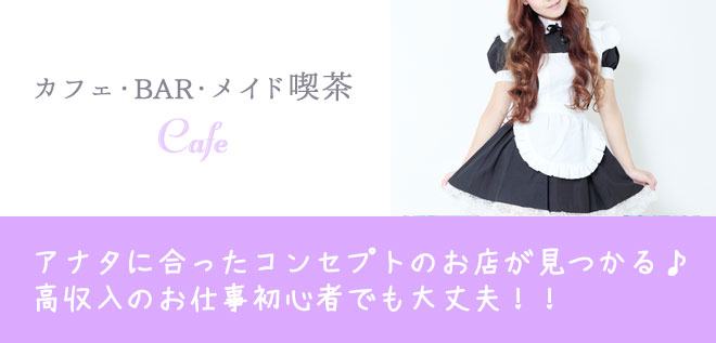
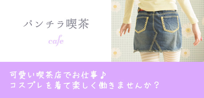
喫茶カフェ系 ▼
平均日給 8500円~35000円以上
| オススメ度 | ★★★★☆ |
|---|---|
| 応募資格 | 18歳以上(高校生不可) |
| 勤務エリア |
【関西】 大阪・京都・兵庫・神戸・滋賀・奈良・和歌山 【関東】 東京・神奈川・横浜・千葉・埼玉・茨木・栃木・群馬 |
| 勤務時間 |
シフト シフト自由で週1日からお仕事可能! 勤務時間 営業時間内から自由に選択可能・応相談 |
| 待遇一覧 | 未経験・初心者OK!/高時給アルバイト/女性の高収入バイト/経験不問/資格不要/資格取得可能!/キャリアアップ/単発・短期・長期OK!/完全自由出勤制/新人保証あり/日給保証あり/時給保証あり/オープニングスタッフ募集/送迎あり/1日体験入店OK!/即日勤務可能/全額日払い/知人バレ・身バレ対策/給与交渉OK!/掛け持ちOK!/週1・月1~OK!/1時間から勤務可能/個人情報保護/専門学生・大学生歓迎!/派遣バイト/深夜/駅チカ/出稼ぎにもおすすめ!/その他 |
平均日給 13000円~30000円以上

| オススメ度 | ★★★★★ |
|---|---|
| 応募資格 | 18歳以上(高校生不可) |
| 勤務エリア |
【関西】 大阪・京都・兵庫・神戸・滋賀・奈良・和歌山 【関東】 東京・神奈川・横浜・千葉・埼玉・茨木・栃木・群馬 |
| 勤務時間 |
シフト シフト自由で週1日からお仕事可能! 勤務時間 営業時間内から自由に選択可能・応相談 |
| 待遇一覧 | 未経験・初心者OK!/高時給アルバイト/女性の高収入バイト/経験不問/資格不要/資格取得可能!/キャリアアップ/単発・短期・長期OK!/完全自由出勤制/新人保証あり/日給保証あり/時給保証あり/オープニングスタッフ募集/送迎あり/1日体験入店OK!/即日勤務可能/全額日払い/知人バレ・身バレ対策/給与交渉OK!/掛け持ちOK!/週1・月1~OK!/1時間から勤務可能/個人情報保護/専門学生・大学生歓迎!/派遣バイト/深夜/駅チカ/出稼ぎにもおすすめ!/その他 |
平均日給 24000円~45000円以上
| オススメ度 | ★★★★☆ |
|---|---|
| 応募資格 | 18歳以上(高校生不可) |
| 勤務エリア |
【関西】 大阪・京都・兵庫・神戸・滋賀・奈良・和歌山 【関東】 東京・神奈川・横浜・千葉・埼玉・茨木・栃木・群馬 |
| 勤務時間 |
シフト シフト自由で週1日からお仕事可能! 勤務時間 営業時間内から自由に選択可能・応相談 |
| 待遇一覧 | 未経験・初心者OK!/高時給アルバイト/女性の高収入バイト/経験不問/資格不要/資格取得可能!/キャリアアップ/単発・短期・長期OK!/完全自由出勤制/新人保証あり/日給保証あり/時給保証あり/オープニングスタッフ募集/送迎あり/1日体験入店OK!/即日勤務可能/全額日払い/知人バレ・身バレ対策/給与交渉OK!/掛け持ちOK!/週1・月1~OK!/1時間から勤務可能/個人情報保護/専門学生・大学生歓迎!/派遣バイト/深夜/駅チカ/出稼ぎにもおすすめ!/その他 |
平均日給 17500円~38000円以上

| オススメ度 | ★★★★☆ |
|---|---|
| 応募資格 | 18歳以上(高校生不可) |
| 勤務エリア |
【関西】 大阪・京都・兵庫・神戸・滋賀・奈良・和歌山 【関東】 東京・神奈川・横浜・千葉・埼玉・茨木・栃木・群馬 |
| 勤務時間 |
シフト シフト自由で週1日からお仕事可能! 勤務時間 営業時間内から自由に選択可能・応相談 |
| 待遇一覧 | 未経験・初心者OK!/高時給アルバイト/女性の高収入バイト/経験不問/資格不要/資格取得可能!/キャリアアップ/単発・短期・長期OK!/完全自由出勤制/新人保証あり/日給保証あり/時給保証あり/オープニングスタッフ募集/送迎あり/1日体験入店OK!/即日勤務可能/全額日払い/知人バレ・身バレ対策/給与交渉OK!/掛け持ちOK!/週1・月1~OK!/1時間から勤務可能/個人情報保護/専門学生・大学生歓迎!/派遣バイト/深夜/駅チカ/出稼ぎにもおすすめ!/その他 |
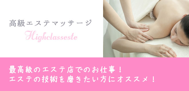
エステマッサージ系 ▼
平均日給 14500円~35000円以上
| オススメ度 | ★★★★★ |
|---|---|
| 応募資格 | 18歳以上(高校生不可) |
| 勤務エリア |
【関西】 大阪・京都・兵庫・神戸・滋賀・奈良・和歌山 【関東】 東京・神奈川・横浜・千葉・埼玉・茨木・栃木・群馬 |
| 勤務時間 |
シフト シフト自由で週1日からお仕事可能! 勤務時間 営業時間内から自由に選択可能・応相談 |
| 待遇一覧 | 未経験・初心者OK!/高時給アルバイト/女性の高収入バイト/経験不問/資格不要/資格取得可能!/キャリアアップ/単発・短期・長期OK!/完全自由出勤制/新人保証あり/日給保証あり/時給保証あり/オープニングスタッフ募集/送迎あり/1日体験入店OK!/即日勤務可能/全額日払い/知人バレ・身バレ対策/給与交渉OK!/掛け持ちOK!/週1・月1~OK!/1時間から勤務可能/個人情報保護/専門学生・大学生歓迎!/派遣バイト/深夜/駅チカ/出稼ぎにもおすすめ!/その他 |
平均日給 18000円~48000円以上
| オススメ度 | ★★★★★ |
|---|---|
| 応募資格 | 18歳以上(高校生不可) |
| 勤務エリア |
【関西】 大阪・京都・兵庫・神戸・滋賀・奈良・和歌山 【関東】 東京・神奈川・横浜・千葉・埼玉・茨木・栃木・群馬 |
| 勤務時間 |
シフト シフト自由で週1日からお仕事可能! 勤務時間 営業時間内から自由に選択可能・応相談 |
| 待遇一覧 | 未経験・初心者OK!/高時給アルバイト/女性の高収入バイト/経験不問/資格不要/資格取得可能!/キャリアアップ/単発・短期・長期OK!/完全自由出勤制/新人保証あり/日給保証あり/時給保証あり/オープニングスタッフ募集/送迎あり/1日体験入店OK!/即日勤務可能/全額日払い/知人バレ・身バレ対策/給与交渉OK!/掛け持ちOK!/週1・月1~OK!/1時間から勤務可能/個人情報保護/専門学生・大学生歓迎!/派遣バイト/深夜/駅チカ/出稼ぎにもおすすめ!/その他 |
平均日給 30000円~50000円以上

| オススメ度 | ★★★★★ |
|---|---|
| 応募資格 | 18歳以上(高校生不可) |
| 勤務エリア |
【関西】 大阪・京都・兵庫・神戸・滋賀・奈良・和歌山 【関東】 東京・神奈川・横浜・千葉・埼玉・茨木・栃木・群馬 |
| 勤務時間 |
シフト シフト自由で週1日からお仕事可能! 勤務時間 営業時間内から自由に選択可能・応相談 |
| 待遇一覧 | 未経験・初心者OK!/高時給アルバイト/女性の高収入バイト/経験不問/資格不要/資格取得可能!/キャリアアップ/単発・短期・長期OK!/完全自由出勤制/新人保証あり/日給保証あり/時給保証あり/オープニングスタッフ募集/送迎あり/1日体験入店OK!/即日勤務可能/全額日払い/知人バレ・身バレ対策/給与交渉OK!/掛け持ちOK!/週1・月1~OK!/1時間から勤務可能/個人情報保護/専門学生・大学生歓迎!/派遣バイト/深夜/駅チカ/出稼ぎにもおすすめ!/その他 |
平均日給 17500円~45000円以上

| オススメ度 | ★★★★☆ |
|---|---|
| 勤務エリア |
【関西】 大阪・京都・兵庫・神戸・滋賀・奈良・和歌山 【関東】 東京・神奈川・横浜・千葉・埼玉・茨木・栃木・群馬 |
| 勤務時間 |
シフト シフト自由で週1日からお仕事可能! 勤務時間 営業時間内から自由に選択可能・応相談 |
| 待遇一覧 | 未経験・初心者OK!/高時給アルバイト/女性の高収入バイト/経験不問/資格不要/資格取得可能!/キャリアアップ/単発・短期・長期OK!/完全自由出勤制/新人保証あり/日給保証あり/時給保証あり/オープニングスタッフ募集/送迎あり/1日体験入店OK!/即日勤務可能/全額日払い/知人バレ・身バレ対策/給与交渉OK!/掛け持ちOK!/週1・月1~OK!/1時間から勤務可能/個人情報保護/専門学生・大学生歓迎!/派遣バイト/深夜/駅チカ/出稼ぎにもおすすめ!/その他 |
平均日給 25000円~50000円以上
| オススメ度 | ★★★★☆ |
|---|---|
| 応募資格 | 18歳以上(高校生不可) |
| 勤務エリア |
【関西】 大阪・京都・兵庫・神戸・滋賀・奈良・和歌山 【関東】 東京・神奈川・横浜・千葉・埼玉・茨木・栃木・群馬 |
| 勤務時間 |
シフト シフト自由で週1日からお仕事可能! 勤務時間 営業時間内から自由に選択可能・応相談 |
| 待遇一覧 | 未経験・初心者OK!/高時給アルバイト/女性の高収入バイト/経験不問/資格不要/資格取得可能!/キャリアアップ/単発・短期・長期OK!/完全自由出勤制/新人保証あり/日給保証あり/時給保証あり/オープニングスタッフ募集/送迎あり/1日体験入店OK!/即日勤務可能/全額日払い/知人バレ・身バレ対策/給与交渉OK!/掛け持ちOK!/週1・月1~OK!/1時間から勤務可能/個人情報保護/専門学生・大学生歓迎!/派遣バイト/深夜/駅チカ/出稼ぎにもおすすめ!/その他 |
平均日給 35000円~80000円以上
| オススメ度 | ★★★★☆ |
|---|---|
| 応募資格 | 18歳以上(高校生不可) |
| 勤務エリア |
【関西】 大阪・京都・兵庫・神戸・滋賀・奈良・和歌山 【関東】 東京・神奈川・横浜・千葉・埼玉・茨木・栃木・群馬 |
| 勤務時間 |
シフト シフト自由で週1日からお仕事可能! 勤務時間 営業時間内から自由に選択可能・応相談 |
| 待遇一覧 | 未経験・初心者OK!/高時給アルバイト/女性の高収入バイト/経験不問/資格不要/資格取得可能!/キャリアアップ/単発・短期・長期OK!/完全自由出勤制/新人保証あり/日給保証あり/時給保証あり/オープニングスタッフ募集/送迎あり/1日体験入店OK!/即日勤務可能/全額日払い/知人バレ・身バレ対策/給与交渉OK!/掛け持ちOK!/週1・月1~OK!/1時間から勤務可能/個人情報保護/専門学生・大学生歓迎!/派遣バイト/深夜/駅チカ/出稼ぎにもおすすめ!/その他 |
平均日給 35000円~70000円以上

| オススメ度 | ★★★★★ |
|---|---|
| 応募資格 | 18歳以上(高校生不可) |
| 勤務エリア |
【関西】 大阪・京都・兵庫・神戸・滋賀・奈良・和歌山 【関東】 東京・神奈川・横浜・千葉・埼玉・茨木・栃木・群馬 |
| 勤務時間 |
シフト シフト自由で週1日からお仕事可能! 勤務時間 営業時間内から自由に選択可能・応相談 |
| 待遇一覧 | 未経験・初心者OK!/高時給アルバイト/女性の高収入バイト/経験不問/資格不要/資格取得可能!/キャリアアップ/単発・短期・長期OK!/完全自由出勤制/新人保証あり/日給保証あり/時給保証あり/オープニングスタッフ募集/送迎あり/1日体験入店OK!/即日勤務可能/全額日払い/知人バレ・身バレ対策/給与交渉OK!/掛け持ちOK!/週1・月1~OK!/1時間から勤務可能/個人情報保護/専門学生・大学生歓迎!/派遣バイト/深夜/駅チカ/出稼ぎにもおすすめ!/その他 |
平均日給 30000円~70000円以上
| オススメ度 | ★★★★★ |
|---|---|
| 応募資格 | 18歳以上(高校生不可) |
| 勤務エリア |
【関西】 大阪・京都・兵庫・神戸・滋賀・奈良・和歌山 【関東】 東京・神奈川・横浜・千葉・埼玉・茨木・栃木・群馬 |
| 勤務時間 |
シフト シフト自由で週1日からお仕事可能! 勤務時間 営業時間内から自由に選択可能・応相談 |
| 待遇一覧 | 未経験・初心者OK!/高時給アルバイト/女性の高収入バイト/経験不問/資格不要/資格取得可能!/キャリアアップ/単発・短期・長期OK!/完全自由出勤制/新人保証あり/日給保証あり/時給保証あり/オープニングスタッフ募集/送迎あり/1日体験入店OK!/即日勤務可能/全額日払い/知人バレ・身バレ対策/給与交渉OK!/掛け持ちOK!/週1・月1~OK!/1時間から勤務可能/個人情報保護/専門学生・大学生歓迎!/派遣バイト/深夜/駅チカ/出稼ぎにもおすすめ!/その他 |
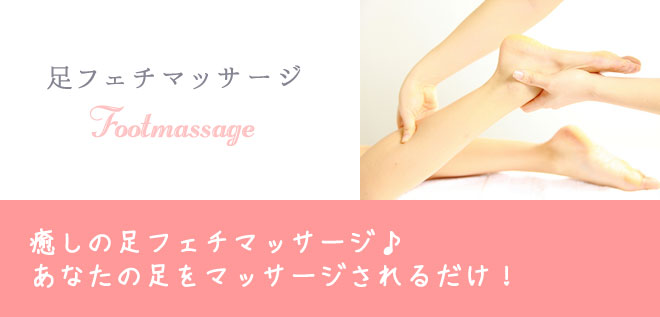
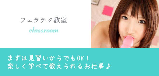
ソフトサービス系 ▼
平均日給 20000円~50000円以上
| オススメ度 | ★★★★☆ |
|---|---|
| 応募資格 | 18歳以上(高校生不可) |
| 勤務エリア |
【関西】 大阪・京都・兵庫・神戸・滋賀・奈良・和歌山 【関東】 東京・神奈川・横浜・千葉・埼玉・茨木・栃木・群馬 |
| 勤務時間 |
シフト シフト自由で週1日からお仕事可能! 勤務時間 営業時間内から自由に選択可能・応相談 |
| 待遇一覧 | 未経験・初心者OK!/高時給アルバイト/女性の高収入バイト/経験不問/資格不要/資格取得可能!/キャリアアップ/単発・短期・長期OK!/完全自由出勤制/新人保証あり/日給保証あり/時給保証あり/オープニングスタッフ募集/送迎あり/1日体験入店OK!/即日勤務可能/全額日払い/知人バレ・身バレ対策/給与交渉OK!/掛け持ちOK!/週1・月1~OK!/1時間から勤務可能/個人情報保護/専門学生・大学生歓迎!/派遣バイト/深夜/駅チカ/出稼ぎにもおすすめ!/その他 |
平均日給 15000円~30000円以上

| オススメ度 | ★★★★☆ |
|---|---|
| 応募資格 | 18歳以上(高校生不可) |
| 勤務エリア |
【関西】 大阪・京都・兵庫・神戸・滋賀・奈良・和歌山 【関東】 東京・神奈川・横浜・千葉・埼玉・茨木・栃木・群馬 |
| 勤務時間 |
シフト シフト自由で週1日からお仕事可能! 勤務時間 営業時間内から自由に選択可能・応相談 |
| 待遇一覧 | 未経験・初心者OK!/高時給アルバイト/女性の高収入バイト/経験不問/資格不要/資格取得可能!/キャリアアップ/単発・短期・長期OK!/完全自由出勤制/新人保証あり/日給保証あり/時給保証あり/オープニングスタッフ募集/送迎あり/1日体験入店OK!/即日勤務可能/全額日払い/知人バレ・身バレ対策/給与交渉OK!/掛け持ちOK!/週1・月1~OK!/1時間から勤務可能/個人情報保護/専門学生・大学生歓迎!/派遣バイト/深夜/駅チカ/出稼ぎにもおすすめ!/その他 |
平均日給 25000円~50000円以上
| オススメ度 | ★★★★★ |
|---|---|
| 応募資格 | 18歳以上(高校生不可) |
| 勤務エリア |
【関西】 大阪・京都・兵庫・神戸・滋賀・奈良・和歌山 【関東】 東京・神奈川・横浜・千葉・埼玉・茨木・栃木・群馬 |
| 勤務時間 |
シフト シフト自由で週1日からお仕事可能! 勤務時間 営業時間内から自由に選択可能・応相談 |
| 待遇一覧 | 未経験・初心者OK!/高時給アルバイト/女性の高収入バイト/経験不問/資格不要/資格取得可能!/キャリアアップ/単発・短期・長期OK!/完全自由出勤制/新人保証あり/日給保証あり/時給保証あり/オープニングスタッフ募集/送迎あり/1日体験入店OK!/即日勤務可能/全額日払い/知人バレ・身バレ対策/給与交渉OK!/掛け持ちOK!/週1・月1~OK!/1時間から勤務可能/個人情報保護/専門学生・大学生歓迎!/派遣バイト/深夜/駅チカ/出稼ぎにもおすすめ!/その他 |
平均日給 35000円~60000円以上

| オススメ度 | ★★★★☆ |
|---|---|
| 応募資格 | 18歳以上(高校生不可) |
| 勤務エリア |
【関西】 大阪・京都・兵庫・神戸・滋賀・奈良・和歌山 【関東】 東京・神奈川・横浜・千葉・埼玉・茨木・栃木・群馬 |
| 勤務時間 |
シフト シフト自由で週1日からお仕事可能! 勤務時間 営業時間内から自由に選択可能・応相談 |
| 待遇一覧 | 未経験・初心者OK!/高時給アルバイト/女性の高収入バイト/経験不問/資格不要/資格取得可能!/キャリアアップ/単発・短期・長期OK!/完全自由出勤制/新人保証あり/日給保証あり/時給保証あり/オープニングスタッフ募集/送迎あり/1日体験入店OK!/即日勤務可能/全額日払い/知人バレ・身バレ対策/給与交渉OK!/掛け持ちOK!/週1・月1~OK!/1時間から勤務可能/個人情報保護/専門学生・大学生歓迎!/派遣バイト/深夜/駅チカ/出稼ぎにもおすすめ!/その他 |
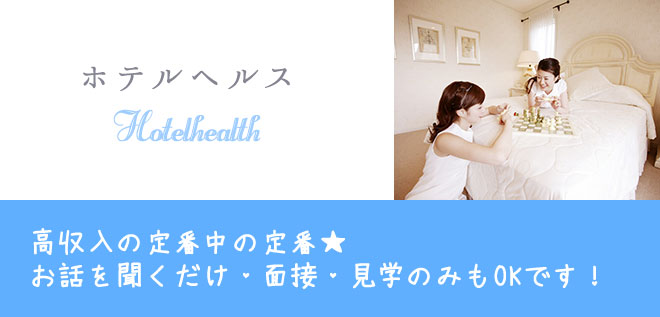
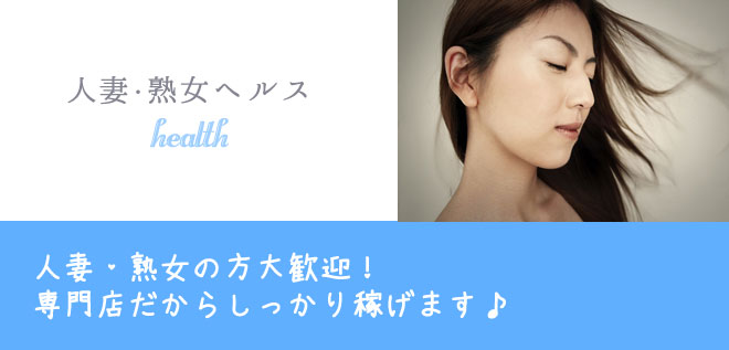
ヘルス系 ▼
平均日給 45000円~10万円以上

| オススメ度 | ★★★★★ |
|---|---|
| 応募資格 | 18歳以上(高校生不可) |
| 勤務エリア |
【関西】 大阪・京都・兵庫・神戸・滋賀・奈良・和歌山 【関東】 東京・神奈川・横浜・千葉・埼玉・茨木・栃木・群馬 |
| 勤務時間 |
シフト シフト自由で週1日からお仕事可能! 勤務時間 営業時間内から自由に選択可能・応相談 |
| 待遇一覧 | 未経験・初心者OK!/高時給アルバイト/女性の高収入バイト/経験不問/資格不要/資格取得可能!/キャリアアップ/単発・短期・長期OK!/完全自由出勤制/新人保証あり/日給保証あり/時給保証あり/オープニングスタッフ募集/送迎あり/1日体験入店OK!/即日勤務可能/全額日払い/知人バレ・身バレ対策/給与交渉OK!/掛け持ちOK!/週1・月1~OK!/1時間から勤務可能/個人情報保護/専門学生・大学生歓迎!/派遣バイト/深夜/駅チカ/出稼ぎにもおすすめ!/その他 |
平均日給 45000円~10万円以上
| オススメ度 | ★★★★★ |
|---|---|
| 応募資格 | 18歳以上(高校生不可) |
| 勤務エリア |
【関西】 大阪・京都・兵庫・神戸・滋賀・奈良・和歌山 【関東】 東京・神奈川・横浜・千葉・埼玉・茨木・栃木・群馬 |
| 勤務時間 |
シフト シフト自由で週1日からお仕事可能! 勤務時間 営業時間内から自由に選択可能・応相談 |
| 待遇一覧 | 未経験・初心者OK!/高時給アルバイト/女性の高収入バイト/経験不問/資格不要/資格取得可能!/キャリアアップ/単発・短期・長期OK!/完全自由出勤制/新人保証あり/日給保証あり/時給保証あり/オープニングスタッフ募集/送迎あり/1日体験入店OK!/即日勤務可能/全額日払い/知人バレ・身バレ対策/給与交渉OK!/掛け持ちOK!/週1・月1~OK!/1時間から勤務可能/個人情報保護/専門学生・大学生歓迎!/派遣バイト/深夜/駅チカ/出稼ぎにもおすすめ!/その他 |
平均日給 35000円~70000円以上
| オススメ度 | ★★★★★ |
|---|---|
| 応募資格 | 18歳以上(高校生不可) |
| 勤務エリア |
【関西】 大阪・京都・兵庫・神戸・滋賀・奈良・和歌山 【関東】 東京・神奈川・横浜・千葉・埼玉・茨木・栃木・群馬 |
| 勤務時間 |
シフト シフト自由で週1日からお仕事可能! 勤務時間 営業時間内から自由に選択可能・応相談 |
| 待遇一覧 | 未経験・初心者OK!/高時給アルバイト/女性の高収入バイト/経験不問/資格不要/資格取得可能!/キャリアアップ/単発・短期・長期OK!/完全自由出勤制/新人保証あり/日給保証あり/時給保証あり/オープニングスタッフ募集/送迎あり/1日体験入店OK!/即日勤務可能/全額日払い/知人バレ・身バレ対策/給与交渉OK!/掛け持ちOK!/週1・月1~OK!/1時間から勤務可能/個人情報保護/専門学生・大学生歓迎!/派遣バイト/深夜/駅チカ/出稼ぎにもおすすめ!/その他 |
平均日給 80000円~25万円以上
| オススメ度 | ★★★★★ |
|---|---|
| 応募資格 | 18歳以上(高校生不可) |
| 勤務エリア |
【関西】 大阪・京都・兵庫・神戸・滋賀・奈良・和歌山 【関東】 東京・神奈川・横浜・千葉・埼玉・茨木・栃木・群馬 |
| 勤務時間 |
シフト シフト自由で週1日からお仕事可能! 勤務時間 営業時間内から自由に選択可能・応相談 |
| 待遇一覧 | 未経験・初心者OK!/高時給アルバイト/女性の高収入バイト/経験不問/資格不要/資格取得可能!/キャリアアップ/単発・短期・長期OK!/完全自由出勤制/新人保証あり/日給保証あり/時給保証あり/オープニングスタッフ募集/送迎あり/1日体験入店OK!/即日勤務可能/全額日払い/知人バレ・身バレ対策/給与交渉OK!/掛け持ちOK!/週1・月1~OK!/1時間から勤務可能/個人情報保護/専門学生・大学生歓迎!/派遣バイト/深夜/駅チカ/出稼ぎにもおすすめ!/その他 |
平均日給 23000円~30000円以上
| オススメ度 | ★★★★☆ |
|---|---|
| 応募資格 | 18歳以上(高校生不可) |
| 勤務エリア |
【関西】 大阪・京都・兵庫・神戸・滋賀・奈良・和歌山 【関東】 東京・神奈川・横浜・千葉・埼玉・茨木・栃木・群馬 |
| 勤務時間 |
シフト シフト自由で週1日からお仕事可能! 勤務時間 営業時間内から自由に選択可能・応相談 |
| 待遇一覧 | 未経験・初心者OK!/高時給アルバイト/女性の高収入バイト/経験不問/資格不要/資格取得可能!/キャリアアップ/単発・短期・長期OK!/完全自由出勤制/新人保証あり/日給保証あり/時給保証あり/オープニングスタッフ募集/送迎あり/1日体験入店OK!/即日勤務可能/全額日払い/知人バレ・身バレ対策/給与交渉OK!/掛け持ちOK!/週1・月1~OK!/1時間から勤務可能/個人情報保護/専門学生・大学生歓迎!/派遣バイト/深夜/駅チカ/出稼ぎにもおすすめ!/その他 |
平均日給 38000円~60000円以上
| オススメ度 | ★★★★☆ |
|---|---|
| 応募資格 | 18歳以上(高校生不可) |
| 勤務エリア |
【関西】 大阪・京都・兵庫・神戸・滋賀・奈良・和歌山 【関東】 東京・神奈川・横浜・千葉・埼玉・茨木・栃木・群馬 |
| 勤務時間 |
シフト シフト自由で週1日からお仕事可能! 勤務時間 営業時間内から自由に選択可能・応相談 |
| 待遇一覧 | 未経験・初心者OK!/高時給アルバイト/女性の高収入バイト/経験不問/資格不要/資格取得可能!/キャリアアップ/単発・短期・長期OK!/完全自由出勤制/新人保証あり/日給保証あり/時給保証あり/オープニングスタッフ募集/送迎あり/1日体験入店OK!/即日勤務可能/全額日払い/知人バレ・身バレ対策/給与交渉OK!/掛け持ちOK!/週1・月1~OK!/1時間から勤務可能/個人情報保護/専門学生・大学生歓迎!/派遣バイト/深夜/駅チカ/出稼ぎにもおすすめ!/その他 |
平均日給 80000円~15万円以上
| オススメ度 | ★★★★☆ |
|---|---|
| 応募資格 | 18歳以上(高校生不可) |
| 勤務エリア |
【関西】 大阪・京都・兵庫・神戸・滋賀・奈良・和歌山 【関東】 東京・神奈川・横浜・千葉・埼玉・茨木・栃木・群馬 |
| 勤務時間 |
シフト シフト自由で週1日からお仕事可能! 勤務時間 営業時間内から自由に選択可能・応相談 |
| 待遇一覧 | 未経験・初心者OK!/高時給アルバイト/女性の高収入バイト/経験不問/資格不要/資格取得可能!/キャリアアップ/単発・短期・長期OK!/完全自由出勤制/新人保証あり/日給保証あり/時給保証あり/オープニングスタッフ募集/送迎あり/1日体験入店OK!/即日勤務可能/全額日払い/知人バレ・身バレ対策/給与交渉OK!/掛け持ちOK!/週1・月1~OK!/1時間から勤務可能/個人情報保護/専門学生・大学生歓迎!/派遣バイト/深夜/駅チカ/出稼ぎにもおすすめ!/その他 |
平均日給 45000円~80000円以上
| オススメ度 | ★★★★☆ |
|---|---|
| 応募資格 | 18歳以上(高校生不可) |
| 勤務エリア |
【関西】 大阪・京都・兵庫・神戸・滋賀・奈良・和歌山 【関東】 東京・神奈川・横浜・千葉・埼玉・茨木・栃木・群馬 |
| 勤務時間 |
シフト シフト自由で週1日からお仕事可能! 勤務時間 営業時間内から自由に選択可能・応相談 |
| 待遇一覧 | 未経験・初心者OK!/高時給アルバイト/女性の高収入バイト/経験不問/資格不要/資格取得可能!/キャリアアップ/単発・短期・長期OK!/完全自由出勤制/新人保証あり/日給保証あり/時給保証あり/オープニングスタッフ募集/送迎あり/1日体験入店OK!/即日勤務可能/全額日払い/知人バレ・身バレ対策/給与交渉OK!/掛け持ちOK!/週1・月1~OK!/1時間から勤務可能/個人情報保護/専門学生・大学生歓迎!/派遣バイト/深夜/駅チカ/出稼ぎにもおすすめ!/その他 |
パーティーサークル系 ▼
平均日給 50000円~10万円以上

| オススメ度 | ★★★★☆ |
|---|---|
| 応募資格 | 18歳以上(高校生不可) |
| 勤務エリア |
【関西】 大阪・京都・兵庫・神戸・滋賀・奈良・和歌山 【関東】 東京・神奈川・横浜・千葉・埼玉・茨木・栃木・群馬 |
| 勤務時間 |
シフト シフト自由で月1日からお仕事可能! 勤務時間 営業時間内から自由に選択可能・応相談 |
| 待遇一覧 | 未経験・初心者OK!/高時給アルバイト/女性の高収入バイト/経験不問/資格不要/資格取得可能!/キャリアアップ/単発・短期・長期OK!/完全自由出勤制/新人保証あり/日給保証あり/時給保証あり/オープニングスタッフ募集/送迎あり/1日体験入店OK!/即日勤務可能/全額日払い/知人バレ・身バレ対策/給与交渉OK!/掛け持ちOK!/週1・月1~OK!/1時間から勤務可能/個人情報保護/専門学生・大学生歓迎!/派遣バイト/深夜/駅チカ/出稼ぎにもおすすめ!/その他 |
平均日給 30000円~50000円以上
| オススメ度 | ★★★★☆ |
|---|---|
| 応募資格 | 18歳以上(高校生不可) |
| 勤務エリア |
【関西】 大阪・京都・兵庫・神戸・滋賀・奈良・和歌山 【関東】 東京・神奈川・横浜・千葉・埼玉・茨木・栃木・群馬 |
| 勤務時間 |
シフト シフト自由で月1日からお仕事可能! 勤務時間 営業時間内から自由に選択可能・応相談 |
| 待遇一覧 | 未経験・初心者OK!/高時給アルバイト/女性の高収入バイト/経験不問/資格不要/資格取得可能!/キャリアアップ/単発・短期・長期OK!/完全自由出勤制/新人保証あり/日給保証あり/時給保証あり/オープニングスタッフ募集/送迎あり/1日体験入店OK!/即日勤務可能/全額日払い/知人バレ・身バレ対策/給与交渉OK!/掛け持ちOK!/週1・月1~OK!/1時間から勤務可能/個人情報保護/専門学生・大学生歓迎!/派遣バイト/深夜/駅チカ/出稼ぎにもおすすめ!/その他 |
平均日給 25000円~50000円以上

| オススメ度 | ★★★★☆ |
|---|---|
| 応募資格 | 18歳以上(高校生不可) |
| 勤務エリア |
【関西】 大阪・京都・兵庫・神戸・滋賀・奈良・和歌山 【関東】 東京・神奈川・横浜・千葉・埼玉・茨木・栃木・群馬 |
| 勤務時間 |
シフト シフト自由で月1日からお仕事可能! 勤務時間 営業時間内から自由に選択可能・応相談 |
| 待遇一覧 | 未経験・初心者OK!/高時給アルバイト/女性の高収入バイト/経験不問/資格不要/資格取得可能!/キャリアアップ/単発・短期・長期OK!/完全自由出勤制/新人保証あり/日給保証あり/時給保証あり/オープニングスタッフ募集/送迎あり/1日体験入店OK!/即日勤務可能/全額日払い/知人バレ・身バレ対策/給与交渉OK!/掛け持ちOK!/週1・月1~OK!/1時間から勤務可能/個人情報保護/専門学生・大学生歓迎!/派遣バイト/深夜/駅チカ/出稼ぎにもおすすめ!/その他 |
平均日給 10000円~30000円以上
| オススメ度 | ★★★★★ |
|---|---|
| 応募資格 | 18歳以上(高校生不可) |
| 勤務エリア |
【関西】 大阪・京都・兵庫・神戸・滋賀・奈良・和歌山 【関東】 東京・神奈川・横浜・千葉・埼玉・茨木・栃木・群馬 |
| 勤務時間 |
シフト 登録依頼制・週1日・月1日でもお仕事可能! 勤務時間 希望時間選択可・応相談 |
| 待遇一覧 | 未経験・初心者OK!/高時給アルバイト/女性の高収入バイト/経験不問/資格不要/資格取得可能!/キャリアアップ/単発・短期・長期OK!/完全自由出勤制/新人保証あり/日給保証あり/時給保証あり/オープニングスタッフ募集/送迎あり/1日体験入店OK!/即日勤務可能/全額日払い/知人バレ・身バレ対策/給与交渉OK!/掛け持ちOK!/週1・月1~OK!/1時間から勤務可能/個人情報保護/専門学生・大学生歓迎!/派遣バイト/深夜/駅チカ/出稼ぎにもおすすめ!/その他 |
【コラム】副業バレないバイト短期日払いアルバイトのおすすめサイト
正業だけではなかなか食べていけない、生活がギリギリという女性も意外に多いのではないでしょうか。まともな企業で働いていてもどうしても収入が少ないというケースはよく聴きますし、今はやりのブラック企業などで働いている場合だと労働に見合った対価を得ていないことも考えられます。 副業してもっと稼ぎたいと思っている方もおられるでしょうが、女性にお勧めの副業だと風俗のナイトワークが挙げられます。風俗のナイトワークならほぼ確実に稼ぐことができますし、週に1回や2回といった働き方もできます。副業には最適ですし、短期間でも可能というお店が多いですからこれもおススメポイントと言えるでしょう。 できれば日払いしてほしい、という方も少なくないでしょうが、多くの風俗店は日払いに対応しています。お店によって対応は異なりますが、基本的に多くの風俗店では日払いや週払いといった支払いに対応しているため安心してください。仕事が終わったら手渡しで給料をもらうことができますし、急に現金が必要になったというシーンでも安心できるのではないでしょうか。アリバイ対策がしっかりしている風俗店ならバレることもありませんからそれについても安心です。
未経験でも大歓迎!気軽に始める副業バイト!

人気カテゴリー
▼おすすめサイト
- 風俗掲示板の噂の口コミ風俗体験談なら【ダンジョウェブ】
- 風俗広告で風俗求人と風俗営業掲載について
- 風俗求人とナイトワーク求人の高収入アルバイト情報【女性高収入ベスト10】
- 【高収入コレクション】女性高収入アルバイト求人
- 高収入バイト風俗求人のナイトワーク求人スカウト
- 【女性高収入バイト副業サポート】女性高収入アルバイト求人高額バイトなら大阪と東京
- 関西の大阪高収入アルバイト求人の京都と神戸【関西アルバイトワーク】
- 【関東アルバイトワーク】関東の東京高収入アルバイト求人の神奈川と埼玉
- 女性高収入アルバイトナイトワーク求人高時給バイト【女性高額収入アルバイトナビ】
- 高【女性の風俗求人口コミweb】収入風俗求人バイト
- 副業バレない高時給バイトなら【副業高収入アルバイトナビ】
- 【モンスタージョブ】女性高収入アルバイト求人
- 女性高収入バイト求人情報の大阪と東京なら【女性高収入バイトサポート】
- 【ハムスタージョブ】副業高時給アルバイト求人で稼げる短期日払いの副業高収入バイト
- 噂の女性高収入アルバイト求人ブログ【セキララ噂の高収入】
- 【一発風俗SEOちゃんねる】風俗広告の営業と求人募集中
- 副業高時給アルバイト風俗求人バイトの【女性高収入求人相談センター】
- 【パンダジョブ】女性高収入アルバイトの副業高時給バイトで風俗求人
